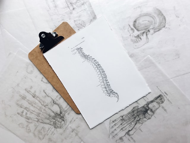
A back and spine MRI is known as a lumbar MRI. It is a scan that uses radio waves, a magnetic field and a computer to generate detailed images of the back area, as well as the spine. It can show organs, soft tissues and skeletal structure as well as abnormal matter, like tumours.
A lumbar MRI is a non-invasive procedure that is commonly used to diagnose many conditions including disk herniation, multiple sclerosis and spinal cord injuries.
MRIs are typically used when a doctor suspects an injury or condition that a typical x-ray isnt able to detect. X-rays are used for ailments that affect dense body parts, so are ideal for things like broken or fractured bones. MRIs, on the other hand, are commonly used for conditions that are harder to identify – or that affect the soft tissue. The reason for this is that MRIs are extremely detailed, so they can show conditions that are of a very small size.
Getting a spine and back MRI is a relatively quick process. The scan itself should only have 10-15 minutes. However, patients should allow more time for the preparation and consultation that will take place before and after the scan.
An MRI scan of the spine and back area can help a doctor discover what the issue is and, consequently, assist them to make a diagnosis and to determine the best treatment plan.

How do back & spine magnetic resonance imaging (MRI) scans work?
A back and spine MRI – or lumbar MRI – works by sending a strong magnetic field to the back area so that all of the atoms in your body to align in the same direction. From here, multiple cross-sectional images are snapped in quick succession.
The images can then be rendered together by a computer program to create a 3-D image of the area. This non-invasive scan can capture bones, cartilage, tendons, ligaments, muscles, and even blood vessels.
A doctor or radiologist will then study these detailed images to help them diagnose the condition that’s causing pain or discomfort.
Why might you need a MRI for your spine or back?
If a doctor suspects an issue within your back and/or spine area that they don’t believe a traditional x-ray would detect, they will often suggest a magnetic resonance imaging scan to confirm their suspicions. Below are some conditions a lumbar spine MRI can detect:
-
Muscle pain
-
Joint damage
-
Sciatica
-
Claudication
-
Spinal cancer
-
Herniated disk
-
Infection of the bone
-
Abscesses
-
Hemorrhage, or bleeding into the spinal cord
-
Encephalomyelitis
-
Multiple sclerosis
If you are experiencing any of the following issues, you may be referred for a lumbar MRI scan:
-
Pain or discomfort anywhere in your back
-
Pain or discomfort in your hips
-
Pain or discomfort in your legs
-
Numbness or weakness in your lower back
-
Loss of movement
-
“Pins and needles” feeling in your legs, toes or feet
-
Cold feet
-
Pressure in your neck, head or back
-
Loss of bladder or bowel control.
-
Difficulty with balance and walking.
How to prepare for your spine or back MRI scan?
A lumbar scan is a non-invasive procedure, so it doesn’t require much special preparation.
However, patients must always remove any metal that they have on their bodies. This includes things like jewellery, a watch, hair pins and removable medical devices. If you have an implanted metal medical device that cannot be removed, like pins and plates for a broken bone, then you should notify the technical before to scan.
Sometimes, a technician will conduct an MRI using a contrast dye. This is done so that the specific parts of the tissue are contrasted from the rest of the body, making it easier to spot certain conditions. A contrast dye is usually administered as a barium meal or intravenously. Usually, patients can still eat and drink before the scan, even if a contrast dye is being used. However, it’s important to always check with your referring doctor.
What To Expect from an MRI scan of your back
During the scan
When you arrive for your scan you will check in with reception like a normal appointment. Once the radiologist is ready for you, you’ll be bought into the MRI room.
Here, you will be given a hospital gown to change into and asked to remove all metal objects if you haven’t already. Once this is done you will be asked to lie on a thin metal bed, and a technician will adjust you so that the scan can correctly capture the area.
You’ll also be fitted with a buzzer and earphones. This will help block out the noise from inside the machine and will allow you to communicate with the technician while inside if required.
MRI machines can be very noisy and are dark and quite small. So, if you suffer from claustrophobia you should let the technician know before the scan. They may decide to sedate you to combat the claustrophobia.
The bed will slowly slide you inside the machine and the images will be taken. The scan itself should only take a few minutes and it’s important that you hold very still. Any movement at all can cause the images to blur.
Once the scan is done, the technician will check that the images are clear and then slide you back out of the MRI machine.
After the scan
Once the MRI exam is over you should be able to continue your day as normal. The exception to this is if you had a sedative for claustrophobia. If so, you may need some recovery time and will need someone to drive you home after the MRI.
In very rare cases, some people can be allergic to MRIs, so if you notice any allergic reactions, like a rash or redness, notify a doctor immediately.
Results from a lumbar MRI usually takes about one week. This gives your physician time to study the images and identify your condition. In some cases, if the condition is serious and needs immediate treatment, your results may be ready sooner.
What are the benefits of an MRI scan of the back?
Lumbar MRIs are an advanced imaging technique that can identify many conditions that a traditional x-ray isn’t able to. Below are some reasons why back and spine MRIs are commonly used.
-
The preciseness of an MRI means that they can pick up very small abnormalities that may not otherwise be evident.
-
MRIs are non-invasive and don’t involve any radiation, meaning they are a very safe procedure.
-
The results are quick, which can mean that treatment can be started quickly.
-
They can identify issues with soft tissues within the back area, which traditional x-rays can’t.
FAQs
Why would a doctor order an MRI for your back or spine?
There are a variety of reasons that a doctor would order an MRI for your back or spine. Some common conditions include MS, Sciatica and general back pain. You can check out the full list above of conditions that can be identified by a spine MRI. Doctors may also order a lumbar MRI to monitor post-surgery recovery.
How long does a back or spine MRI take?
The scan itself should only take about 15 minutes. However, patients should allow 60-90 minutes for the whole appointment, to allow for preparation and consultation time.
What does a back or spine MRI show?
A lumbar MRI shows detailed 3-d images of the spine and back area; it clearly shows organs, soft tissues and skeletal structure as well as abnormal matter, like tumours.
Is a back or spine MRI uncomfortable?
Because a back or spine MRI is a non-invasive procedure, there shouldn’t be any discomfort. However, sometimes a doctor will need to position you in a certain way so that the scan can correctly capture all of the required areas. This positioning may be slightly uncomfortable. However, if you ever feel any discomfort or pain at all, you should always tell your technician straight away.
Does your head go in for a back or spine MRI?
Sometimes. If you’re entering the machine head first, then your head will be inside the MRI machine. Sometimes, patients are able to enter an MRI machine foot first, and in this case, they may be able to keep their heads outside of the MRI machine.
Can I drink water before a back or spine MRI?
Usually, yes. However, it’s important to always check with your referring physician prior to the appointment, as some doctors may have different requirements.
Will a doctor automatically recommend an MRI if I have back or spine issues?
No. A doctor will speak to you about your symptoms and conduct a physical exam. If they believe you have a condition that can be diagnosed through an MRI, they will refer you for a scan.


