What is a
chest x ray?
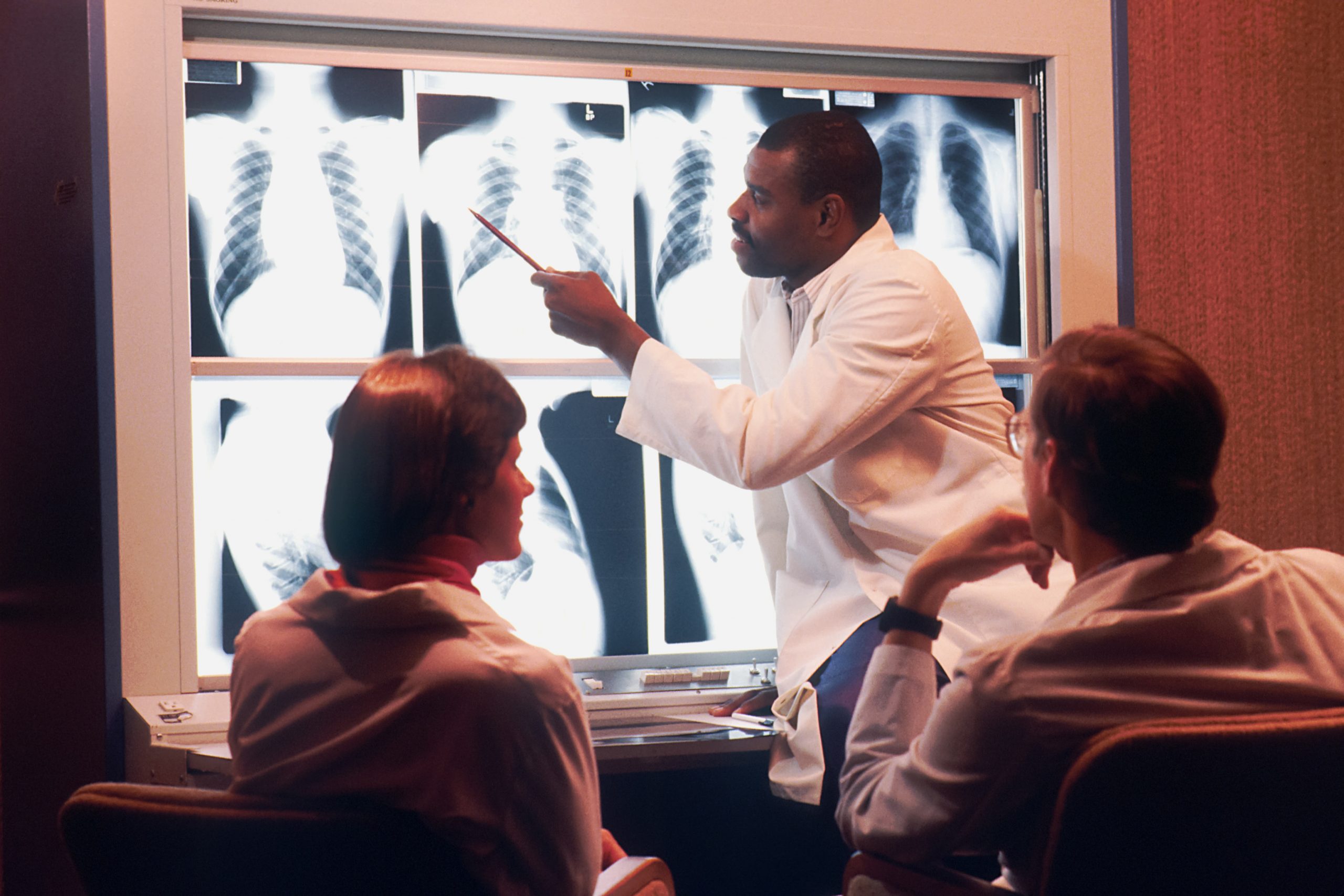
A chest x ray is essentially a machine that can project an image of your chest onto a screen that generates a printed image on x ray film that a doctor or radiologist can read to assess your health.
An x ray is taken by a radiologist, whose profession is to specialise in the imaging of human anatomy and technologists have specific training in X-ray testing.
As well as performing x rays, radiologists are also trained in using other imaging technology that takes scans like ultrasounds, MRI’s and CT scans.
If a patient feels unwell and a doctor predicts the condition has something to do with their chest, the doctor will usually order a chest x ray for the patient.
What conditions can a chest x ray detect?
There are many conditions that can be a result of a problem with the chest area. That’s why chest x rays are a good way for a doctor to determine the cause of the health problem. Some conditions that may affect the chest are as follows:
Pneumonia
Pneumonia is an injection that inflames the air sacs of the lungs and can be caused by a virus, bacteria, fungi or other germs. The condition can be fatal if it leads to serious complications. A chest x ray can identify if a patient has pneumonia. The radiologist will look for white spots in the lungs (called infiltrates) that identify an infection.
Tuberculosis
Tuberculosis is an infectious disease that can be spread by droplets that travel through coughing and sneezing. Tuberculosis isn’t very common, but, it’s resistant to a lot of treatments, so it can be very dangerous. A chest x ray can detect abnormalities that may indicate tuberculosis.
Lung cancer
Lung cancer is the uncontrolled growth of abnormal cells in one or both lungs. A radiologist will look for a white-grey mass on a chest x ray as an indication of lung cancer.
Breast cancer
Like lung cancer, breast cancer is a disease where abnormal cells in the breasts grow out of control. A chest x ray can help to detect whether breast cancer has spread to the lungs too.
Enlarged heart
An enlarged heart is usually indicative of a heart valve problem or heart disease. There are other serious ailments that have an enlarged heart as a symptom. A chest x ray will identify an enlarged heart and a doctor can do more tests to find out the cause.
Blocked arteries
If arteries are blocked blood flow to the heart will be decreased and a completely blocked coronary artery will cause a heart attack. A chest x ray can find changes in the size, shape and position of the major blood vessels and arteries.
Rib cage injuries
Rib cage injuries can come about from sudden accidents, like a fall or a car crash. Ribcage injuries can be a variety of things, but include broken ribs, fractures, internal bruises and torn cartilage. Chest x rays will show what damage is done to the ribs and a doctor can recommend the best treatment.
Congestive heart failure
Congestive heart failure is when the heart doesn’t pump blood as well as it should. Chest x rays can show changes or problems in your lungs that stem from heart problems which can indicate congestive heart failure.
Why should I get chest x ray?
How do I prepare for a chest x ray?
What happens when I go for a chest x ray?
Once all metal objects are removed and you are wearing the x ray gown you can enter the x ray room.
To take the front image of your chest the radiologist will instruct you to stand with your chest against the metal plate of the X-ray machine and your hands on your hips.
The radiologist will enter a connecting room where she or he can take the x ray images.
You’ll then need to turn around so that the x ray technician can take x ray images on the side of your chest. For this, you will be instructed to stand with your side against the metal plate of the x ray machine and your arms in the air.
It’s important that you hold very still while the x ray technician is taking the chest x ray. Even breathing in and out can blur the image.
Once the radiologist is happy with the chest x rays, you’ll be able to leave the room and will be finished. Chest x rays generally only take a few minutes.
Ready to make an appointment?
If you’d like to find out more information about a specific treatment, to book a consultation or to make an appointment to see a dr, you can get in touch with our friendly staff at our clinic here.
FAQ’s
Are there any risks involved with getting chest x rays?
Radiologists are medical doctors that have spent years studying and training for their position. Their level of skill will ensure that you are no put in any danger when in the x ray room. General chest x rays are completed every day and are a common way to diagnose chest conditions.
Humans are actually exposed to radiation in their everyday life. The amount of radiation that is emitted during a normal chest x ray is very small and is the equivalent amount of radiation an average human (who didn’t have an x ray) would be exposed to over a 10 day period.
How long does it take to get results from an x ray?
Chest x rays produce instant images onto x ray film. So the x rays are actually available immediately. However, a radiologist will need time to review an x ray and identify the problem. Depending on the urgency of the condition or problem, it could take a few days to get results or you could have them instantly.


