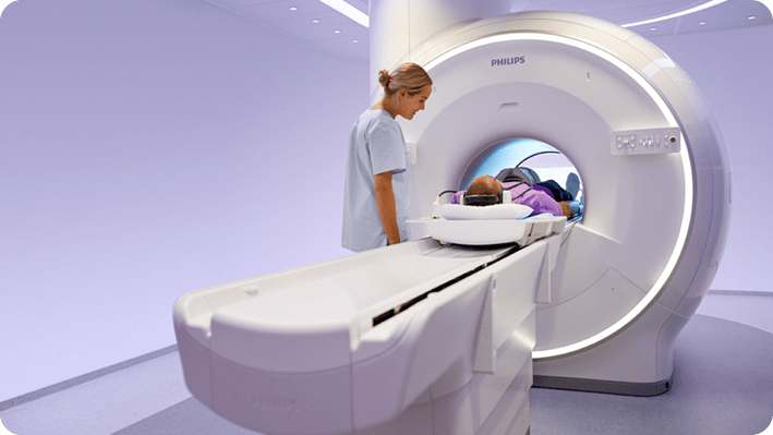
An ankle injury is one of the most common type of sports injuries. But, while ankle injuries are often found in athletes, they can also be caused by everyday activities. Furthermore, ankle conditions can extend further than just injuries – things like tumours, infections, necrosis and arthritis can all develop in the foot and lower limb area.
An ankle MRI is a useful tool to detect and diagnose ailments of the ankle. It can detect things like tendon, ligament and cartilage injuries, as well as broken bones, tumours and infections.
An ankle MRI has the ability to pick up a much more detailed image than an x-ray, and take multiple cross-sectional pictures of the ankle, called slices. These high-quality images can be joined together to create a 3D representation of the ankle region.
An ankle MRI can help a doctor discover what the issue is and, consequently, assist them to make a diagnosis and help them to determine the best treatment plan.

How do ankle MRI scans work?
An ankle MRI is a magnetic resonance imaging test that shows a detailed picture of the ankle area. This type of scan is more details than an x-ray, and will show things that an x-ray can’t, like ligaments, tendons and muscles.
An MRI generates a large number of cross-sectional images in quick succession, these axial images can then can be joined together after the scan is finished through the use of 3D rendering. The 3D representation of the area helps doctors to see things that they wouldn’t be able to on a flat image and helps them identify even very small issues.
The process of getting an Ankle MRI is usually quick and easy, and patients should leave the test feeling completely normal. However, there is a different type of MRI, which is called a contrast MRI. This type of test is rarely used in ankle scans, but some physicians may choose to. It involves administrating a contrast dye to a patient, usually through IV or a barium meal, and this dye helps physicians to identify problems more clearly. This type of scan will likely take longer than a normal MRI.
Another factor that can make an MRI slightly more complex is when a patient is administered a sedative for the scan. This is usually only done when a patient is very claustrophobic, as the testing machine is dark and narrow. If a patient is sedated for the test, they will need some recovery time set aside for after the test.
Why might you need an ankle MRI?
If a doctor suspects an issue with a ligament or tendon in the ankle, they will almost always suggest an MRI to confirm their suspicions. However, ankle MRIs can detect far more than just ligament and tendon injuries or tears. Below are some conditions an ankle MRI can detect:
-
Arthritis
-
Ankle fractures
-
Stress fractures
-
Partial tear of the tendon or ligament
-
Injury of the anterior talofibular ligament, posterior talofibular ligament and/or lateral collateral ligament
-
Tears of the peroneal tendons or flexor hallucis longus tendon
-
Lesions or Tumours
-
Lateral ankle sprains
-
Posterior tibial tendon abnormalties
-
Tarsal tunnel syndrome
-
Inflammation
-
Infection
-
Achilles tendon injury
-
Plantar fasciitis
-
Posterior tibial tendon dysfunction
-
Avascular necrosis
If you are experiencing any of the following issues, you may require an ankle MRI.
-
Swelling in ankle
-
Tenderness in ankle
-
Bruising in ankle
-
Pain in ankle
-
An injury that affected the ankle area
-
Immobility of the ankle
-
Stiffness of the ankle
How to prepare for your ankle MRI scan
An ankle MRI is a non-invasive procedure, so there isn’t much preparation that’s required. However, metal objects shouldn’t be worn into MRIs, so it’s a good idea to remove these before your appointment so that the test can be completed quicker.
Patients should remove metal items like a watch, jewellery and hairpins. Some makeup and hairspray contain metal particles too, so it’s recommended to skip using these products until after the test. If you have a surgically implanted metal device, like a hearing aid or metal pins from an accident, then you should tell your doctor about these prior to the scan.
If you have claustrophobia, you should inform your referring doctor about your condition. If they recommend a sedative for the test, then they may be some associated rules, like not being able to eat or drink for a certain time before the test.
What To Expect from your ankle MRI scan
During the scan
When you arrive for your abdominal x-ray, you will check with the receptionist like a normal doctor’s appointment. When it’s your time for the scan, you will be taken to the x-ray room where you’ll find a medical table and an x-ray machine hanging over the top.
With an abdominal x ray, you will almost always be given a hospital gown to change into. Once you’re dressed in the gown, you’ll be asked to sit on an exam table. A technician will help to positive you so that you’re lying correctly so that the x ray machine will capture the correct areas. Sometimes they’ll put a lead mat over other areas of your body that don’t need to be captured in the photo/s.
After this, the technician or radiologist will head into a separate room or an area that’s partitioned off. They will then capture the images and it’s important that you stay very still during this time. Even the slightest movement can create a blur in the photos.
Depending on your specific issue, the technician may come back and position your differently a few times, so that they can capture images of different areas.
Once the photos are taken, the technician will usually ask you to wait a few minutes while they check that the x-rays are all clear. If so, you’ll be able to change back into your regular clothes and continue about your day as normal.
After the scan
An abdominal x ray is a non-invasive procedure, so there generally won’t’ be anything special you need to do once the x ray is completed. However, in very rare cases, a patient can have an allergic reaction to an x-ray. So, if you notice any symptoms of an allergy at all, like hives or a rash, then you should contact a doctor immediately.
The images from abdominal x-rays are available immediately. However, in most cases, a doctor will need some time to analyse them and diagnose the problem. Once this is done, the doctor’s office will contact you for a follow-up appointment to discuss your results and their findings. This will usually be a day or two after the appointment. If your condition is urgent, a doctor may prioritise your results and have them ready immediately.
What are the benefits of ankle magnetic resonance imaging (MRI)?
Ankle MRIs are an advanced imaging technique that can identify many conditions that a traditional x-ray isn’t able to. Below are some reasons why ankle scans are commonly used.
-
-
The preciseness of an MRI means that they are able to pick up very small abnormalities that may not otherwise be evident
-
They can identify issues with soft tissues within the wrist, which traditional x-rays can’t
-
MRIs are non-invasive and don’t involve any radiation, meaning they are a very safe procedure.
-
The results are quick, which can mean that treatment can be started quickly
-
FAQs
Why would a doctor order an ankle MRI?
In order to get an ankle MRI, a patient will have to be referred from a doctor or surgeon. This means that a physician will look at your ankle and listen to your symptoms, and if they belive an MRI may be able to help diagnose the condition, they will refer the patient for one. Some common reasons that patients are referred for an ankle MRI include Achilles tendon injury, Plantar fasciitis, and ligament injuries.
How long does an ankle MRI take?
An ankle MRI is relatively quick, and the scan itself should only take about 10-15 minutes. However, patients should set aside at least an hour for their appointment. This will cover the waiting and preparation time, as well as the time required for the scan itself.
Is an ankle MRI uncomfortable?
An ankle MRI is a non-invasive procedure. So, a patient should feel uncomfortable during the scan. However, if the patient has an injured ankle, and it must be adjusted at a particular angle in order to capture the images properly, then they may feel minor discomfort. If you feel pain – or any discomfort – at any time during the MRI, you should always let the radiologist of techincian know.
Will a doctor automatically recommend an MRI if I have acute ankle pain?
No. Some conditions can be diagnosed through an x-ray or a physical exam. A doctor will consult with you to listen to symptoms, and then they will conduct a physical exam so that they can get an idea of what area is affected. If they suspect that an MRI will be able to diagnose the condition, they will refer you for one.


