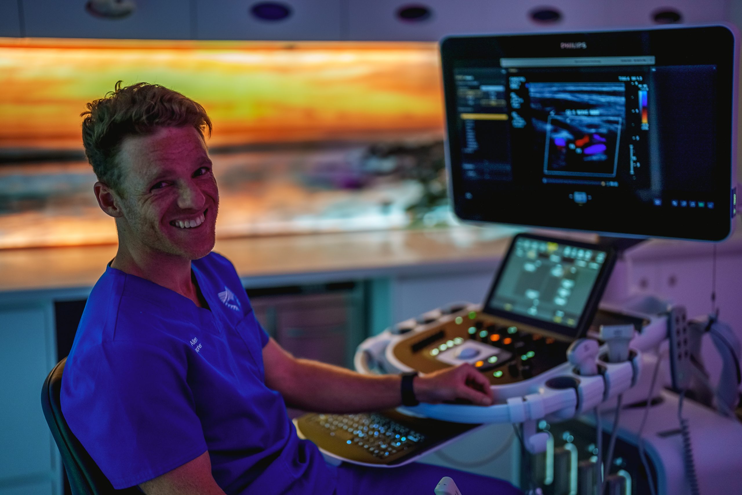
Living with endometriosis can be overwhelming and frustrating, from the pain to swinging hormone levels to missed periods. It’s understandable if you feel lost in the world of medical terminology or unsure of what to do next. One potential diagnostic tool that could help bring clarity is an endometriosis ultrasound—but what exactly is it? What kind of preparation should you make? How does the process work? All essential questions will be addressed below so you can be better informed. Take a closer look for an overview that will provide helpful guidance towards understanding and navigating a successful endometriosis ultrasound procedure!

What Is Endometriosis Ultrasound?
Endometriosis ultrasounds use soundwaves to get a detailed view of your body to determine if endometrial tissue is found in places other than the uterus. Endometriosis can affect different organs, including the ovaries, bladder and intestines. During the process, your healthcare professional will use specialized transducers. This will detect changes in sound wave reflections which could indicate endometrial tissue. You’ll most likely be presented with a visual onscreen of images from the ultrasound. This way, you can understand more about the condition and how it’s affecting your body.
Purpose of an Endometriosis Ultrasound
Like other medical procedures, an endometriosis ultrasound is used to gain a lot of information about a person’s body without the need for an invasive, high-risk procedure. Knowing the extent of endometriosis, its location and how much it has progressed can help physicians devise a tailored treatment plan. For some women, this knowledge can be life-changing. But here are some of the purposes of an endometriosis ultrasound:
Checking for the Presence of Endometriosis
Ultrasound scan is a valuable tool that can help diagnose endometriosis. This is used to check for the presence of endometriosis in women who may be exhibiting signs of the condition. During the ultrasound, medical professionals will be looking for telltale signs, such as cysts or abnormal developments in the pelvic and abdominal area. A successful ultrasound scan can provide valuable insight into a woman’s health and can be vital to getting an accurate diagnosis and proper treatment for this condition.
Assessing the Severity of the Disease
Endometriosis ultrasounds can be an incredibly useful tool for diagnosing endometriosis and evaluating its severity. With the scan, a medical professional is able to detect areas of chronic pelvic pain and deep infiltrating endometriosis to get an idea of the overall severity of the condition. This can help determine treatment options. For instance, mild cases of endometriosis might respond best to over-the-counter medications, whereas more severe cases may require surgery.
Evaluating the Symptoms
Evaluating suspected endometriosis through an ultrasound might seem like a daunting task, but it is an important one. During the procedure, the radiologist looks for cysts that could be causing pain or other symptoms and scans deeply to determine if there are any signs of deeply infiltrating endometriosis. Examining these suspected areas of the pelvic anatomy can help you, and your doctor assess which treatments will work best to relieve your symptoms and maybe even slow down disease progression.
Monitoring Disease Progression
Monitoring pelvic endometriosis with ultrasound is a critical part of long-term treatment as it can help to identify any changes or new areas of tissue growth in the fallopian tubes. This early detection is important so that adjustments can be made to ensure that the condition isn’t progressing too quickly and that treatment is effective. While ultrasound imaging isn’t the only way to track endometriosis progression, it is a reliable tool for keeping a close eye on the condition over time.
Guiding the Treatment
Women with suspected endometriosis can benefit significantly from an endometriosis ultrasound. This scan can identify any growth of endometrial tissue and even detect if there are cysts present. Based on the results, treatment options like surgery or medication may be recommended to help women manage their symptoms. Importantly, an endometriosis ultrasound can provide crucial information that will assist women in making informed decisions about how to best treat their condition and potentially achieve a better outcome.
Preparation for an Endometriosis Ultrasound
Because of this, some medical professionals recommend that you perform a mild bowel preparation prior to the ultrasound. If you have a history of severe endometriosis or major bowel pain during your periods, this procedure may be recommended for you. In general, this is done when you have severe endometriosis. This involves consuming a gentle laxative the night before the ultrasound to clear your system. It is highly recommended that you consume two full glasses of water around half an hour before your scheduled scan time. Make sure that you remember to bring the completed referral form with you so that the scan is done correctly and efficiently. Having the necessary details on hand will help the radiologist make a quicker and more accurate diagnosis.
Process of an Endometriosis Ultrasound
When you arrive with a full bladder, the sonographer will ask you to lie down on the bed and expose your stomach. At this point, a towel will be placed into your pants to keep them from moving around. Following this, a sonographer will apply a gel that is based on water to your lower abdominal region (just below the belly button). This gel helps to improve the contact that the probe has with your skin, thus, it’s important to use it. After that, the technologist will slide the probe around your lower abdomen while taking photos of the pelvic region.
Afterwards, you will be instructed to vacate your bladder, change into a gown, and return to the examination room. Then the sonographer will perform a transvaginal ultrasound on the patient. You will have a specialized probe inserted into your vagina. This probe will have a sterile cover and will be used. It will be moved in a variety of ways so that diagnostic images can be obtained, which will assist in determining the cause of your pain. The last step of the procedure is for the sonographer to take measurements and then provide a report about the findings.
Risks Involved in an Endometriosis Ultrasound
There are some risks associated with getting an endometriosis ultrasound, but don’t worry – it’s usually not too painful. You should be aware that vaginal exams can occasionally cause bleeding for up to 24 hours afterwards. This is normal and shouldn’t last any longer than this. However, if you experience prolonged bleeding get in touch with your doctor straight away! One thing worth bearing in mind when having the scan done is that depending on where exactly the bowels lie, the technician may run into difficulties viewing all areas affected by endometriosis due to the angle of the ultrasound. So make sure to ask questions if something seems unclear during the examination.
Outcomes From an Endometriosis Ultrasound
An endometriosis ultrasound can provide an invaluable diagnosis — and sometimes even peace of mind. Although at times, it may be a bit uncomfortable, the outcome is well worth it. After the doctor reviews the results of your ultrasound, they can tell you whether or not you have endometriosis. They will then suggest potential treatments depending on their severity. It may involve medication or even surgery. That’s why it’s essential to listen carefully to all of the guidance given during your appointment. If followed, this advice could make an incredible difference in how you cope with the condition. It’s wise to heed your doctor’s advice to help manage your condition, as it could make a huge difference in how you feel day-to-day.
When To See a Doctor About the Results of Your Scan?
Within the next 48 hours, your doctor will have access to the whole report of the scan. Navigating when to see a doctor about the results of your scan can be tricky. We always recommend speaking with a certified healthcare professional as soon as you feel like something is off. In fact, if your scan reveals something that could be classified as clinically serious, our team will come and talk to you about it on the same day. On the other hand, there are many findings from scans that are normal and do not require conversation. Therefore, we encourage patients to trust their judgment. If something doesn’t seem right, speak up! It’s better to be safe than sorry when it comes to your health.
Conclusion
Endometriosis ultrasound is a necessary procedure that can help diagnose and manage endometriosis. This offers a fast, non-invasive way to get detailed information about the presence of lesions in the uterus and other organs. It helps to reduce risk by allowing for early detection of any serious conditions associated with endometriosis. With its low risks, minimal preparation requirements, and quick results, this type of scan is becoming increasingly popular among women looking to take charge of their healthcare decisions. By considering the purpose, preparation, process, risks and outcomes involved in an endometriosis ultrasound above, you can make an informed decision about how to best manage your condition.


