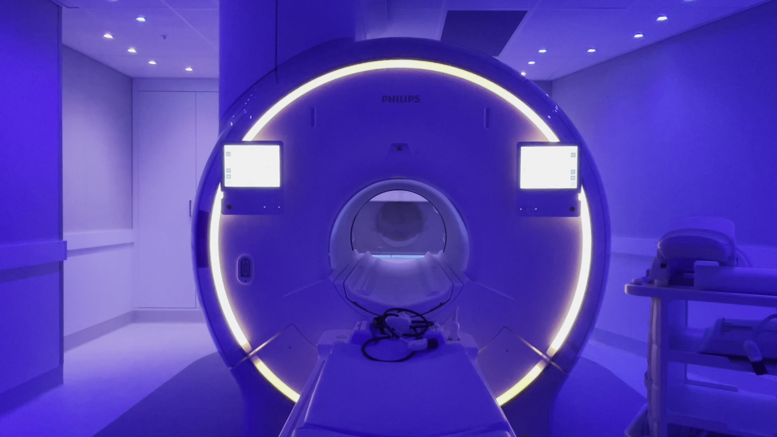
Magnetic Resonance Imaging is an imaging strategy that utilises radio waves or magnetic fields to create clear images of the body’s internal structures. It is also known in examining ovaries and even on other part of our reproductive system.
But why would we need an ovarian MRI? There’s a few reason on why professionals may utilise this kind of MRI scan. Let’s look at the information for an ovarian MRI and what to expect during the procedure.
Magnetic Resonance Imaging is an imaging strategy that utilises radio waves or magnetic fields to create clear images of the body’s internal structures.
But why would we need an ovarian MRI? There’s a few reason on why professionals may utilise this kind of MRI scan. Let’s look at the information for an ovarian MRI and what to expect during the procedure.

What Is an Ovarian MRI and Why Is It Important?
This non-surgical procedure uses magnetic fields and radio waves to create a detailed ovary scan. It is a safer imaging method for reproductive organs than X-rays due to the reason of involvement of radiation.
The significance of an ovarian MRI cannot be overrated, particularly when it comes to detecting ovarian cancer and other diseases like germ cell tumors or pelvic inflammatory disease.
So when examining for malignant ovarian tumors, MRIs are essential in confirming the mass, location and size of the said tumour.
Therefore, early detection of ovarian malignancy can increase the success rate of treatment of conditions affecting the female pelvis or ovaries.
How To Prepare for Your Ovarian MRI Scan
-
Consult your doctor: Discuss your health conditions, allergies, or pregnancy with your healthcare provider. Also, inform them about implants, pacemakers, artificial joints, or cochlear implants.
-
Dress comfortably: You must change into a hospital gown for the procedure. It’s advised to wear loose, comfortable clothes that are easy to change out of.
-
Avoid metallic items: Because the MRI uses strong magnetic fields, removing metallic objects before the scanning is essential. The safety and accurateness of the MRI scan are important, so it’s necessary to inform your healthcare provider about any metallic implants or devices in your body.
-
Fasting: MRI scans often require the patient to have an empty stomach to ensure the best imaging findings. It is done to avoid any interference that food or liquids might cause in the MRI process.
-
Arrive early: Try to arrive for at least 30 minutes before your scheduled appointment. It will give you sufficient time to complete the necessary paperwork and preparation for the scan.
What to Expect During an Ovarian MRI Scan
Once you’re all set for the ovarian MRI, you might wonder what the procedure will be like. Let’s walk through what you can expect during your scan:
Entering the MRI Room and Positioning for the Scan
During the procedure, you’ll be accompanied into the MRI room, which has a large machine with a bed that slides inside a tunnel-like structure. The room is usually kept cool, so you might be offered a blanket for comfort.
It may seem intimidating, but it’s designed to be as comfortable as possible. Your safety is the practitioners’ priority; the room has equipment such as an intercom. There is even a panic button in case of any discomfort or emergency.
Positioning for the Scan
After entering the MRI room, the technologist will help position you onto the bed. Generally, you will be asked to lie down on your back. Also, pillows and cushions might make you comfortable and keep you still during the scan.
They’ll then ensure all metal has been removed and you’re set correctly for the best potential visions. Once everything is in place, the technologist will slide the bed into the machine, where the magnetic field is designed for the imaging.
Use of a Contrast Agent
Often, an ovarian MRI scan may involve a contrast agent. This dye helps emphasise certain areas in the image, providing a clearer view of your ovaries.
Before the scan, this contrast agent is administered through an intravenous (IV) line inserted into your arms. You might feel a cool sensation as injected, but it’s completely normal. After the scan, the agent is safely eradicated via your kidneys.
The Scanning Process
Once you’re comfortably positioned and prepared, the table will slide into the MRI apparatus. The machine can be loud, casting, humming or thumping noises throughout the scanning process — this is normal and part of how the machine operates.
Often, you’ll be provided headphones or earplugs to help block out the noise. You can use this time to relax and even hear some music. Remember that remaining still is essential for acquiring clear images, so do your best to keep your body relaxed and stationary.
Duration of the Scan
An ovarian MRI scan commonly takes 30 to 60 minutes to complete. However, the duration can differ depending on the specific details of the scan. The procedure might prolonged if a contrast agent is utilised.
The technologist overseeing your scan will keep you informed about how long you have left and will check in on you periodically to ensure your comfort.
Knowing the Different Types of Images Produced by an Ovarian MRI Scan
The MRI scan of the ovaries generates different types of images, each providing unique insights into the health of your ovaries.
- T1-weighted images: These images are often applied to detect peritoneal implants and identify areas of fat and water inside the body. In a T1-weighted image, the fat appears bright, while the water appears dark. These imaging features provide a clear view of the structures and enable the radiologist to identify any abnormalities.
- T2-weighted images: T2-weighted images, on the other hand, make water or fluid appear bright and fat appear dark. It is beneficial in identifying cysts in the ovaries, as they are fluid-filled and will stand out in these images.
- Diffusion-weighted images (DWI): It’s beneficial in distinguishing benign from malignant (cancerous) ovarian masses. Since cancer cells are typically densely packed, the diffusion of water is restricted and thus appears differently on a DWI.
- Post-contrast images: It’s taken after the injection of the contrast agent. The contrast dye improves the clarity of the blood vessels, and it can differentiate normal and abnormal tissues. It can also highlight any malignant tumours in the ovaries, aiding in the findings of ovarian cancer.
Each type of image is vital in getting an accurate diagnosis of your ovarian health. Your healthcare provider can make differential diagnoses accurately and plan effective treatments through this scan.
Understanding the Results of an Ovarian MRI Scan
After your ovarian MRI scan, a radiologist – will review the collected images. They will find signs of abnormalities, such as cystic lesions, tumours or structural changes in the normal ovaries.
The radiologist will gather a detailed report based on their understanding of the images; this outcome will then be forwarded to your healthcare provider. Rest assured, every image is carefully reviewed and compiled meticulously to ensure the most accurate assessment.
During your follow-up appointment, your healthcare provider will consult the results with you. They will inform you about the findings and the possible interventions.
Remember, having concerns about your results is entirely normal. Don’t hesitate to ask your health provider for clarification or further explanation. Understanding the results of your ovarian scan is vital for your overall health.
Benefits and Risks Associated With Undergoing an Ovarian MRI Scan
To decide on any medical procedure, it’s essential to understand its benefits and risks.
Benefits of Undergoing an Ovarian MRI Scan:
-
Non-invasive and painless: MRI scans don’t require surgery and are completely painless, making them more comfortable for patients.
-
Highly detailed images: MRI scans offer detailed images of the ovaries, making it easier for doctors to notice abnormalities such as tumours or cysts.
-
No radiation: Unlike CT scans, MRI scans do not use radiation, reducing the potential risks of radiation exposure.
Risks Associated With Undergoing an Ovarian MRI Scan:
-
Allergic reactions: Though rare, some may be allergic to the contrast agent or dye used in MRI scans.
-
Claustrophobia: Claustrophobic patients might experience discomfort during the MRI scan as the procedure involves lying in a narrow, enclosed space.
-
Incompatibility with specific medical devices: Some medical instruments, such as pacemakers or cochlear implants, might not be compatible with MRI machines.
A Deeper Look into Abnormal Findings in an Ovarian MRI
When learning the outcomes of an MRI, familiarising yourself with the possibilities of what could be discovered is essential. Abnormalities can range from benign to malignant; some common abnormal findings in an ovarian or pelvic MRI scan include the following:
-
Ovarian cysts: This cystic lesion can form in the ovaries during menstrual cycles. While most ovarian cysts are benign and often go unnoticed, they can sometimes grow large, causing discomfort and pain. Due to their fluid content, they’ll appear as bright on the T2-weighted images.
-
Ovarian masses/tumours: Refer to any abnormal growth in the ovaries, which can be benign or malignant. Further assessment is necessary to determine the classification and differential diagnosis. Also, to determine the nature of ovarian lesions and tumours, plan appropriate treatment.
-
Polycystic ovary syndrome (PCOS): In PCOS, multiple small cysts grow in your ovaries. The MRI might show the ovaries slightly bigger than normal, with multiple small cysts.
-
Ovarian cancer: The most severe of all possible findings is ovarian cancer and malignant lesions. It can be life-threatening if not seen and treated earlier. MRI scans are useful in detecting ovarian cancer at an early stage, improving survival rates.
-
Endometriomas: These are cysts caused by endometriosis, where tissue identical to the uterus lining grows outside of it. Endometriomas can be specified by their distinct ‘shading’ effect on T2-weighted images.
Final Thoughts
MRI of ovaries is a device for detecting and diagnosing ovarian conditions or pelvic organs. Your medical specialist may recommend an Ovarian MRI if you are experiencing signs such as pelvic pain or irregular menstrual cycles.
Even though MRIs carry some risks, the benefits of the procedure is greater than the concern and help you improve. Also, remember to continuously consult with your healthcare provider if you have any worries or questions about your ovarian health.


