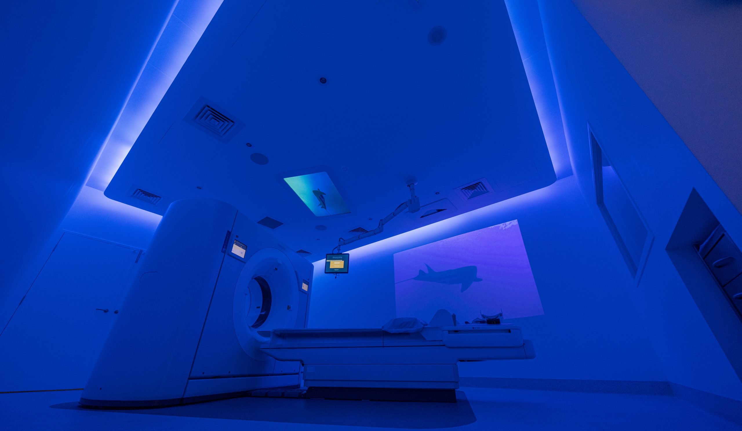
Pulmonary fibrosis (PF) is a condition that concerns the lungs, making it difficult for individuals to ventilate. If the disease is found in the human body this will lead to respiratory failure. One of the key ways in which this condition is diagnosed and observed is through CT scans.
One of the key ways in which this condition is diagnosed and monitored is through CT scans. In the case of IPF, CT scans can show areas of scarring in the lungs and help doctors determine the extent and progression of the fibrotic lung disease.
But what do these CT scans exactly show? How can CT Scan aid in the diagnosis of PF? Here are some key information and an understanding of Pulmonary Fibrosis Through CT Scans:

To What Extent Does Pulmonary Fibrosis Affect the Lungs?
Long-term scarring and thickening of lung tissue can lead to interstitial lung disease, such as idiopathic interstitial pneumonias or nonspecific interstitial pneumonia. This thickening or scarring, known as fibrosis, makes it more difficult for your lungs to function as they should. As the condition advances, you might lack breath, even when resting.
Our lungs are designed to be elastic, stretching and contracting with every breath we take. In a healthy lung, oxygen easily passes into your bloodstream, producing essential energy our bodies need. However, with pulmonary fibrosis, this process is severely disrupted.
As fibrosis occurs, the tissue’s elasticity reduces, making it challenging for the lungs to move and for oxygen to pass into the bloodstream. Consequently, the brain and other organs become starved of the oxygen they need to function optimally.
The Role of CT Scans in Diagnosing Pulmonary Fibrosis
CT scan of pulmonary fibrosis are important in diagnosing PF, allowing you to detect and intervene in premature stages. Doctors can identify any changes that may occur in pulmonary fibrosis by providing images of the lungs.
These scans can show how much damage has occurred with the combination of blood tests and X-rays. Additionally, CT scans can help doctors specify the level of the disease and monitor its response to treatment.
In conclusion, CT scan of pulmonary fibrosis are vital in interpreting and monitoring pulmonary fibrosis. By providing images of the lungs, doctors can identify and track any changes or damage that may occur in the lungs.
Results of CT Scan in Pulmonary Fibrosis
In CT scan results for a patient with Pulmonary Fibrosis and idiopathic pulmonary fibrosis, you might come across various findings:
Honeycombing: This term refers to small cystic areas with well-defined walls of fibrotic tissue, typical in the advanced stages of pulmonary fibrosis. It’s called so because it resembles a beehive.
Ground-glass opacity (GGO): refers to areas that appear hazy on the CT scan. The affected regions allow some lung markings to be seen through them, akin to looking through frosted or ground glass.
Reticulation: This term is used to describe a web of lines. In the context of CT scans, it refers to a network of small linear opacities, which usually represents the thickening of the interlobular septa.
Traction bronchiectasis: This appears when the surrounding fibrotic tissue pulls and distorts the bronchi, causing them to appear larger and irregular.
Volume loss: Volume loss or shrinkage of the lung tissue can occur as the fibrosis progresses, indicated by moving the heart, blood vessels, or airways towards the affected part.
Consolidation: This term is employed when a lung region or lung area becomes filled with liquid instead of air, making it appear more solid on the Computerised Tomography Scan.
Common Treatments for Pulmonary Fibrosis
Treating pulmonary fibrosis or fibrotic lung diseases primarily involves managing the symptoms, improving the patient’s quality life and slowing the disease’s progression. Here are some commonly used treatments for pulmonary fibrosis:
Medication
Drugs like pirfenidone and nintedanib can slow the progression of pulmonary fibrosis in some people. Immunosuppressants may control and reduce inflammation that could lead to chronic hypersensitivity pneumonitis or fibrosis.
Oxygen Therapy
It’s a simple and effective way to ensure your body gets oxygen. It is pretty common in managing pulmonary fibrosis. This treatment involves breathing in oxygen through a mask or prongs in your nose.
Pulmonary Rehabilitation
Pulmonary Rehabilitation, often called “Pulm Rehab”, is the gym for your lungs. It’s led by health professionals who guide you through pulmonary function tests, provide tips and help improve your overall well-being.
Palliative Care
The main goal of this approach is to enhance the patients’ and families’ quality life. It addresses physical, intellectual and emotional issues. It may involve managing shortness of breathing and providing relief from distressing symptoms.
Lung Transplant For Severe Pulmonary Fibrosis
In severe cases of pulmonary fibrosis, where other treatments are no longer effective, doctors may suggest a lung transplant. The patient has to have their damaged lungs replaced with healthy ones from a donor during this difficult medical procedure.
Understanding the Different Types of CT Scans Used to Assess Pulmonary Fibrosis
When it comes to CT scans for Pulmonary Fibrosis, High Resolution Computed Tomography (HRCT) and standard Computed Tomography scans are the two main types of diagnosis without being subjected to surgical lung biopsy.
The HRCT is particularly efficient in diagnosing lung fibrosis like pulmonary fibrosis; providing highly detailed images of the lung tissues. It can capture thinner slices of the lung, allowing doctors to spot the most minor abnormalities and changes in lung tissue.
On the other hand, a standard CT scan, though not as detailed as an HRCT, is still a valuable tool in diagnosing and monitoring pulmonary fibrosis. It is frequently utilised by clinicians when they require a wider picture of the lungs and surrounding structures since it can capture larger sections of the lung.
Both types of CT scans have their own unique roles in the fight against pulmonary fibrosis. The choice between HRCT and standard CT scans depends on various factors, including the individual’s condition, symptoms, and the doctor’s assessment.
The Benefits of CT Scanning for Diagnosing PF and Tracking Its Progression Over Time
CT Scanning presents numerous benefits for diagnosing Pulmonary Fibrosis (PF) and tracking its progression over time:
Early Detection
Early pulmonary fibrosis (PF) detection is almost impossible, but with CT scans, it becomes a possibility. To Diagnose pulmonary fibrosis it needs questioning and CT scans for your lungs, whether an HRCT or a standard CT can reveal signs of PF even before symptoms show.
They can identify the scarring and thickening of lung tissues, often the first indicators of this disease. It also means that treatment can begin earlier, potentially delaying the progression of the disease like usual interstitial pneumonia and improving the quality of life for those affected.
Detailed Visualisation
CT scans are often likened to peering into a window that grants an insider’s view of the body. They offer detailed images and cross-sectional views of the lung tissues, painting a comprehensive picture of what’s happening inside.
This visualisation capability proves invaluable in developing tailored treatment plans, assessing their effectiveness, and adjusting them as needed. It’s like having a roadmap for navigating the complex journey of managing pulmonary fibrosis, informing every step.
Monitoring Disease Progression
It is essential to monitor the conditions and manage the progress of pulmonary fibrosis. With Computerised Tomography scans or CT scans it provide a dynamic way to constantly track the development of pulmonary fibrosis.
By comparing the past and latest results, the radiologist can track the ongoing increase or decrease in inflammation or lung scarring. This continuous monitoring aids in adjusting treatments and symptom management in intervening in diseases.
Guiding Treatment Plans
CT scans are essential for the diagnosis, tracking, and assistance of therapy plans for pulmonary fibrosis. These scans enable medical professionals to develop treatment plans based on each patient’s unique situation by providing images of the lungs.
CT scans guide the medical team in formulating the most practical treatment approach for patients with pulmonary fibrosis. Suggesting the need for medication changes or identifying potential complications, CT scans contribute significantly to the decision-making process.
Non-Invasive Diagnosis
In the context of pulmonary fibrosis or idiopathic pulmonary fibrosis, one of the significant advantages of CT scans is their non-invasive nature. CT scans do not involve any incisions or physical intrusion into the body. It means no stitches, no extensive recovery time, and significantly less discomfort.
This ease of diagnosis not only minimises physical discomfort for patients but also reduces anxiety and stress associated with invasive diagnostic procedures. Computerised Tomography scans provide a less complicated and more straightforward route in identifying and treating pulmonary fibrosis.
Final Thoughts
CT scan plays an important role in managing and detecting pulmonary fibrosis; it also aids in diagnosis helping monitor disease progression and treatment plans.
With their non-invasive diagnosis, it offers a less intimidating detection and effective management of the diseases. Additionally, numerous treatments such as rehab or medicines, are available to manage the disease progression.
So, if you are experiencing any symptoms or have risk factors for pulmonary fibrosis, talk to your healthcare provider about getting a CT scan and exploring treatment options.


