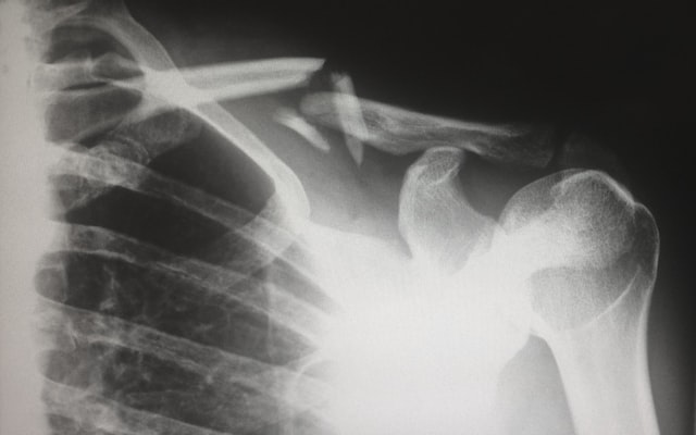
Joints are complex structures that are made up of bone, muscles, synovium, cartilage, and ligaments. When these structures are injured, it can be painful, difficult or impossible to raise the arm up.
Standard X-rays don’t show soft tissues, like ligaments and tendons, whereas MRIs do. This is why MRI scans are very helpful with detecting problems in the shoulder area.
An MRI scan of the shoulder can help a doctor discover what the issue is and, consequently, assist them to make a diagnosis and to determine the best treatment plan.

How do shoulder MRI scans work?
These images are cross-sectional images, which means the images produced are sliced through the middle of the structure. Because a number of images are taken in quick succession, a computer program can be used the stack the images up and make them appear three-dimensional, through the use of 3D rendering. This preciseness can help a doctor detect small conditions that may otherwise be missed.
Sometimes, a physician will require a patient to have a contrast MRI scan of the shoulder. This means that a contrast agent (like a dye) will be used and it will be administered intravenously or the patient will be asked to take it as a barium meal. A contrast agent can improve the clarity of the body’s internal structures, helping a radiologist to make a diagnosis.
Unlike X-rays, an MRI doesn’t use radiation. Instead, it harnesses radiowaves from data that already exist in the body, meaning it won’t cause chemical change or damage to tissue.
Why might you need a shoulder MRI?
Often, a standard x-ray will be ordered first, to see if the problem can be detected. If not, an MRI can show more detail and may be able to identify the issue. While MRI’s are very helpful in detecting issues within the soft tissues, they can show a variety of other conditions, too.
If you are experiencing any of the following issues, you may require a shoulder MRI.
- Unexplained swelling or pain in the shoulder
- Lack of mobility in the shoulder
- Pain when arms are lifted
- Shoulder injuries from an accident or fall
- Persistent pain that doesn’t improve over time
- A popping sound in the shoulder when arms are raised or moved
- If an abnormality is noticed on an x-ray
- If an x-ray can’t detect the reason for shoulder discomfort or pain
Below are a number of conditions that a shoulder CT scan can help diagnose.
- Dislocation of the shoulder joint
- Rotator cuff tears or injuries
- Arthritis in the shoulder
- Infections
- Tissue damage
- Torn ligaments or tendons
- Bone tumours
- Inflammation in the shoulder
- Degenerative joint disease
- Sprains
- Bone fractures or breaks
How to prepare for your shoulder MRI scan
Once you’ve received your referral, you’ll need to go ahead and book an appointment for your magnetic resonance imaging scan.
After you’re booked in for your scan, there’s not much you need to do in way of preparation.
Some doctors will conduct a contrast scan, and this may require you to fast for a given amount of time. However, this isn’t always the case, so it’s best to check with your referring physician about whether you’re able to eat or drink before your appointment.
Prior to attending your scan, you should remove all metal objects, like earrings, piercings and hair pins. Metal objects can obstruct the scan which can cause issues when the technician is reading them. If you have any internal metal implants that can’t be removed, like a pacemaker or hearing aid, then you should let your radiologist know about it prior to the scan.
What To Expect from your shoulder MRI scan
During the scan
At your appointment, you will be given a hospital gown to change into. If you are having a contrast scan the doctor will administer this agent through an IV or ask you to consume it as a barium meal.
The technician will then fit you with some earplugs or headphones to drown out some of the noise that the MRI scanner makes. They will also give you a buzzer, which, when pressed, will let you speak to the technician during the scan.
After this, the technician will ask you to lie on a narrow metal table, and will adjust your body so that the scanner can take clear pictures of your shoulder. The table will slowly slide you inside the MRI scanner. The machine is a small metal shape tube and is narrow, so if you experience claustrophobia you should let your doctor know prior to the scan. If they see fit, they may offer you a sedative to help calm you.
Once inside, you must ensure that you hold completely still, any movement at all can blue the images. Don’t be surprised or off-put by the loud sound during the scan, the machine is very noisy.
While the scan is happening, a patient shouldn’t feel anything major. Sometimes, patients feel a little warm, while this isn’t usually something to worry about, it’s always a good idea to tell your physician if you do experience this.
Once the shoulder MRI scan is completed, the patient will be slid out of the machine and be allowed to change back into their original clothes.
After the scan
MRI scans are a non-invasive process, so a patient should feel normal after the scan is completed. The exception to this is if a sedative was administered for claustrophobia-related reasons. If this is the case, a patient will require a recovery period and will need someone to drive them home.
Sometimes, a patient will notice that they feel slightly nauseous, or have local pain in the region of the scan. While this is usually injury-related, a medical professional should always be notified if you feel any discomfort.
In rare cases, a patient may have an allergic reaction to an MRI scan. So, if you notice anything that may signify an allergy, like a rash, you should let a doctor know immediately.
What are the benefits of shoulder magnetic resonance imaging (MRI)?
- They can identify issues with soft tissues within the shoulder, which traditional x-rays can’t
- The preciseness of an MRI means that they are able to pick up very small abnormalities that may not otherwise be evident
- MRIs are non-invasive and don’t involve any radiation, meaning they are a very safe procedure.
- The results are quick, which can mean that treatment can be started quickly
FAQs
How long does a shoulder MRI take?
A shoulder MRI will normally take between 30 – 60 minutes, but in some cases, it can take up to two hours. This time includes the preparation, a patient will generally only spend a few minutes inside the machine for the actual scan.
When do you need an MRI for shoulder pain?
An MRI will be issued by a doctor. Usually, a doctor will conduct a physical exam and then order a standard x-ray. If the x-ray doesn’t show the problem, a doctor will usually order an MRI. However, If a doctor suspects that the symptoms show an issue that won’t be picked up in a standard x ray, they will skip this step and go ahead and order an MRI after the physical exam.


