Wrist pain can stop us in our tracks – the simple everyday tasks that were once effortless now become a challenge. It’s easy to shrug it off and tell ourselves we just need to wait for it to pass, but persistent or sudden wrist pain is not something you want to ignore.
If you’re experiencing heightened discomfort symptoms, certain tests are available that could diagnose your underlying issue – and one such test is an MRI scan of your wrist.
To help you, we’ll explain why getting an MRI is important if you’re dealing with wrist pain, what sort of results you should expect afterwards, along with potential treatment options depending on your diagnosis.

What is a Wrist MRI Scan?
A wrist MRI scan is a sort of medical imaging examination that uses magnetic fields and radio waves to provide precise images of your wrist’s soft tissues, bones, tendons, and ligaments. It’s used to diagnose conditions such as carpal tunnel syndrome, tendonitis, fractures, etc. The test is non-invasive and doesn’t involve radiation, making it a safe and adequate way to diagnose issues.
This type of scan can provide better images than a traditional X-ray, as it can distinguish between the various tissues in your wrist. It uses magnetic fields to align the protons in your body’s cells and then sends pulses of radio waves through the area being scanned. This creates detailed pictures that doctors can use to diagnose conditions.
How Do Wrist MRI Work?
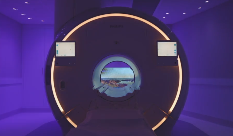
In a wrist MRI scan, you’ll be asked to place your wrist in a special scanner. The machine uses magnetic fields and radio waves to take images of the innards of your wrist. Your body will stay still during the procedure so that the machine can capture clear images.
The MRI scan is painless, but you may feel discomfort as your wrist is placed inside the machine. The technician will monitor your comfort level throughout the procedure and adjust the settings if needed.
Once the images are taken, they’ll be sent to a doctor or radiologist for review. The specialist will be able to review the images and determine if any damage or disease is present in the wrist. In some cases, they may recommend further testing or treatment based on their findings.
Why Might You Need a Wrist MRI Scan?
If you possess any of the following indications and symptoms, you should consider having a wrist MRI scan:
• Severe pain or tenderness in one area of your wrist
• A restricted range of motion in your wrist
• Pain that radiates to your fingers
• Swelling or discolouration around the affected area
• Instability in your wrist joint
• Hearing a popping or grinding noise when you move your wrist
• Pain on the outside of your wrist that worsens with activity
• Weak grip strength or difficulty gripping objects
• A feeling like your wrist is going to give out or become dislocated
• Numbness, tingling, or a burning sensation in one area of your wrist
These are all common signs and symptoms that could mean you have an underlying condition in your wrist joint. If you’ve experienced any of these, it might be time to consider getting a Wrist MRI scan.
How to Prepare for Your Wrist MRI Scan?
Preparation for a wrist MRI scan is relatively straightforward and usually does not require any special steps.
You should wear comfy clothing that you can easily remove when necessary. Metal objects, such as jewellery or buttons, may interfere with imaging and should be removed before your scan. Inform your doctor if you have metallic implants like a pacemaker or metal clips on your blood vessels. You may be requested to switch to a hospital gown for the MRI scan.
Sometimes, your doctor may instruct you to stop taking particular medications before the scan. This is because certain drugs can interfere with MRI scans and the results of the scan. It is necessary that you follow your doctor’s instructions regarding any necessary medication adjustments carefully.
What to Expect From Your Wrist MRI Scan?
If you’ve been informed that you need a wrist MRI scan, it can be daunting. You may not understand what to expect or if the scan is necessary. But don’t worry. It’s natural to be nervous before any type of medical procedure, and understanding what will happen during your scan will help put your mind at ease.
During a wrist MRI scan, you’ll be positioned on the exam table, and your arm will be placed inside an enclosed tunnel that contains the imaging equipment. You may feel pressure as the imaging coil is placed around your wrist, but it should not be uncomfortable. The radiographer will then move away from the room to control the MRI scanner from the next room.
Your wrist will then be scanned, and images of your carpal bones, distal radioulnar joint, ligaments, and tendon sheath will be taken. The technologist may need to move your arm during the scan for different angles or in order to focus on a particular area. You should try to remain still throughout the procedure to make the images clear.
When the scan is ended, you will be able to leave the room. The images from your wrist MRI scan will then be transmitted to a radiologist for interpretation and diagnosis. Depending on what the radiologist finds, they may recommend further tests or treatment options if necessary.
After having a wrist MRI scan, you must follow all of your doctor’s or medical staff’s directions. This may include wearing a splint or brace to support the wrist and help reduce pain, taking medications, or undergoing physical therapy. These actions will aid the recovery process and provide long-term relief from any discomfort caused by your injury or ailment.
What Are the Benefits of Wrist Magnetic Resonance Imaging (MRI)?
A wrist MRI scan is one of the most reliable procedures for identifying and treating wrist injuries or disorders. There are many benefits to getting a wrist MRI, all of which can help determine the severity of an injury and find possible solutions to healing. Below are 5 of the benefits associated with having a wrist MRI.
Help to Diagnose a Wide Variety of Conditions
Dealing with certain health conditions can be tough, especially when figuring out what’s happening inside your body. Thankfully, innovative diagnostic imaging options are now accessible, including wrist MRI.
This diagnostic imaging tool is good at finding injuries and diseases like scaphoid waist fractures, tendonitis, transverse carpal ligament tears, and arthritis.
Plus, it is super reliable when other imaging techniques like ultrasound or X-ray fail to provide a definitive diagnosis. And the best part is wrist MRI can provide doctors with a more thorough and precise view of the problem area.
Non-invasive and Does Not Use Ionising Radiation
A wrist MRI scan is a non-invasive technology used to evaluate the structure of the wrists and hands. Unlike X-rays or computed tomography (CT) scans, it does not use ionising radiation, making it safer for medical imaging.
This makes it ideal for imaging patients more frequently, such as athletes who may be at risk of repetitive hand and wrist injuries. With a wrist MRI scan, doctors can monitor any changes occurring over time without exposing the patient to unnecessary radiation.
Provide Clear Images of the Bones and Soft Tissues
A wrist MRI scan is an effective way to look at your wrist anatomy and soft tissues. The images produced by a wrist MRI scan can help you and your doctor diagnose any conditions or injuries that may be causing discomfort in your wrist.
This allows for detailed visualisation of the wrist’s bones, ulnar collateral ligament, extensor pollicis longus tendon, ulnar nerve, median nerve and muscles. With a wrist MRI scan, you can see the location and orientation of each bone in your wrist. You can also see the relationship between anatomical structures of scaphoid and lunate bones and how they work together to allow for movement.
Painless and Generally Well Tolerated by Patients
If you’re worried about having an MRI of the wrist, you’ll be relieved to know that most people regard it as a painless treatment. While some may experience mild discomfort from their arm positioning, this is short-lived and isn’t severe enough to require pain medication. So there’s no need to worry about excruciating pain during the scan.
Additionally, patients often report that the entire procedure is quick and straightforward. So there’s no excuse to delay getting the answers you need about your wrist health with this non-invasive and tolerable imaging technology.
Used in Conjunction With Other Imaging Modalities
Regarding wrist conditions, doctors need the whole picture to diagnose. That’s why they use a cool technique called ‘used in conjunction with other imaging modalities, combining a wrist MRI with other scans like an X-ray or CT scan. It’s like putting together a puzzle but with no missing pieces.
By doing this, doctors better understand what’s happening inside your wrist and can find the best ways to treat you. So if you get a wrist MRI, don’t be surprised if more scans are involved. It’s all about getting you back on track for recovery.


