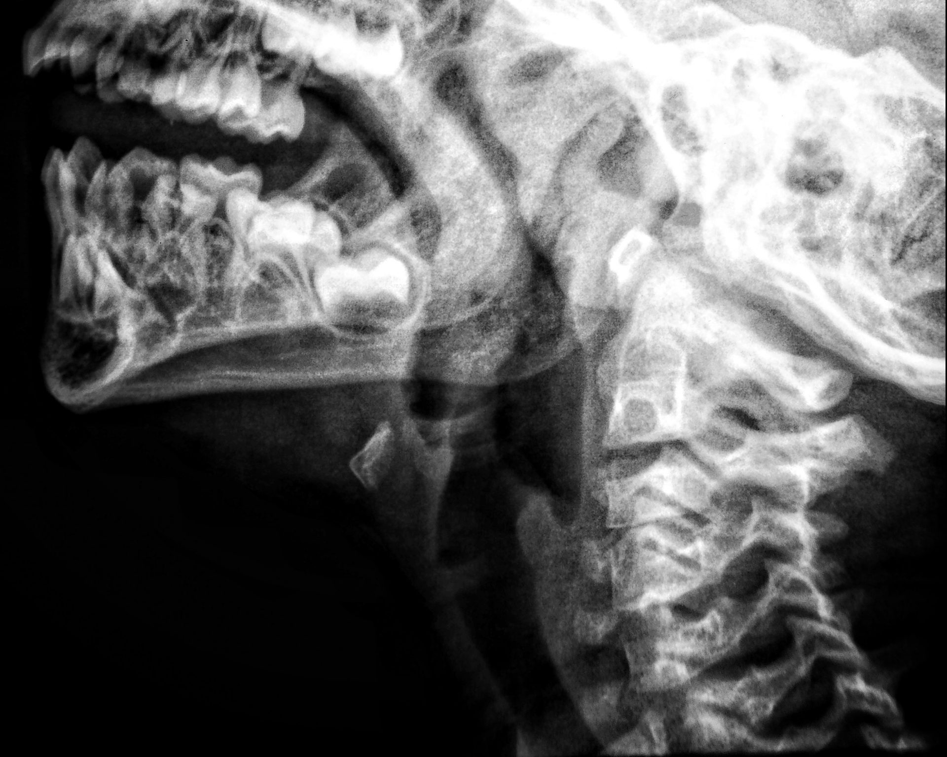
What is a neck x ray and how does it work?
Neck X rays are scans that are conducted by a technician that help a doctor or radiologist diagnose conditions or abnormalities in the area. This imaging procedure is commonly used to detect broken or fractured bones, but can also capture images of the tissue.
Neck x-rays are commonly known as a cervical spine x-ray or cervical vertebrae x ray because they look at the cervical vertebrae. The cervical vertebrae is made up of seven neck bones, including the first vertebrae of the thoracic spine, that surround and protect the top section of the spinal cord.
X-rays work by casting a small amount of radiation and electromagnetic energy beams into the area being scanned, in this case, the neck. These black and white images are then reflected onto a screen and printed onto x-ray film. Hard matter, like bones, blocks the x-ray beam, so they show up as white on the film. Soft tissue allows the beam to pass through, so they show up in a lighter colour. Because of this, if there is a break or fracture in the bone, the beam will seep through, showing a black line and this is how doctors determine whether the bone is broken.
X-rays are almost always ordered after a consultation with a doctor. If a doctor suspects a broken or fractured bone, they will order an x-ray. If they suspect an issue with soft tissue then they will likely recommend an MRI. MRI’s are more detailed scans that create images that are viewed as cross sections or “slices”.
Why might you need a neck x-ray?
As mentioned above, a neck x-ray or cervical spine x ray is usually conducted by a doctors order. So, a patient should have already seen a general physician about their symptoms. If a patient is experiencing any of the following issues, a doctor may order a cervical vertebrae x-ray.
-
Neck, shoulder, upper back, or arm pain
-
Tingling in the arm or hand
-
Numbness in the arm or hand
-
Weakness of the arm or hand
-
Fractures or breaks in the cervical vertebrae
-
Dislocation of the joints between the vertebrae
-
If you’ve been involved in an injury that affects the pelvis
Below are a number of conditions that a neck x-ray can help diagnose.
-
Fractured bones
-
Broken bones
-
Arthritis
-
Spondylolisthesis
-
Degeneration of the disks
-
Kyphosis
-
Scoliosis
-
Congenital abnormalities
How to prepare for your neck x-ray
A neck x-ray doesn’t’ require any special preparation. However, before doing the scan a technician will ask you to remove any metal from your body, so it’s a good idea to take these off before going to your appointment. This is because metal objects will obstruct the imaging test and will block photos, so a technician won’t be able to see the bones and soft tissues in that area.
These metal objects include piercings, jewellery, eyeglasses and a watch. If you have metal implanted in your body permanently, like a pace maker or pins and plates for a broken bone, then you should tell your technician about this before the exam. Some makeup and hair spray contains metal particles, too, so you should remove this before doing the scan.
What to expect from your neck x-ray
During the scan
When you arrive at your scan, once you’ve checked in with the receptionist, you’ll be taken to an x-ray room. Here, the technician can answer any questions you may have about the x-ray. After this, you’ll be given a hospital gown to change into for the scan.
Then, a technician will ask you to lay on an x ray table, below the hanging x-ray machine. They will assist to position you so that the x-ray can capture the correct areas of your body. Sometimes, they’ll place a led blanket over areas that they don’t’ need to capture, as this will block the radiation.
After this, the technician or radiologist will head into another room, or an area that’s partitioned off. They will then take the photos and it’s very important that you stays very still at this time. Even the smallest movement can create blurry images.
If a technician requires multiple positions in order to properly detect your condition, they may move or adjust you so they can photograph other areas.
Once this is done, the technician will probably ask you to wait a few moments while they check if all the images are clear. If so, the scan is finished and you can continue about your day as usual.
After the scan
A neck x-ray is a non invasive procedure, so there’s no after care that’s required and you should feel completely normal. In very rare cases patients can have allergic reactions to an x-ray, so if you noticed any allergy symptoms, like a rash or hives, then you should notify a doctor immediately.
The results for a neck x-ray usually take about 1-2 days to come back. If your doctor detects an urgent condition, they may prioritise your results and get them back sooner. Once the doctor has checked the x-rays and anaylsed them, they will organise a follow up appointment with you to discuss their findings.
What are the benefits of a neck x-ray?
Below are some reasons why spine x-rays are commonly used.
-
X rays are non invasive and painless.
-
X rays are very quick, in many cases your results will be available within a few days.
-
They are very useful in detecting a number of conditions, and are an easy and effective way to determine if you have a broken bone or fracture.
-
They are relatively cheap, in comparison to a CT scan or a magnetic resonance imaging test.
FAQs
Are neck x-rays harmful to my health?
Contrary to what many people believe, x-rays are actually have very low radiation exposure. People actually already have radiation exposure in their every day life. In fact, the radiation exposure in a hand x-ray is the equivalent amount of radiation an average human (who didn’t have an x ray) would be exposed to over a 10 day period.
Radiologists are medical doctors that have spent years studying and training for their position. Their level of skill will ensure that you are no put in any danger when in the x ray room. Neck x rays are completed every day and are a common way to diagnose chest conditions.
How long does a neck x ray take?
This will depend on how many positions your technician requires you to be in, in order to capture adequate images. The actual x-ray process only takes a couple of minutes per position. However, you should set aside 30-45 minutes for an appointment, to include time to speak to the technician and change into the hospital gown.
Can a neck x-ray show a pinched nerve?
The answer to this isn’t clear cut. Technically, an x-ray doesn’t show nerves, so a damaged nerve won’t show on a neck x-ray. However, the x-ray will show the positioning of bones, and this may be able to indicate whether a nerve is damaged or pinched. To officially diagnose a pinched nerve, another test like an MRI will be required.
Can a neck x-ray show a herniated disc?
Much liked a pinched nerve, a standard x-ray isn’t able to show a herniated disc. However, it will show the outline of your spine, which can help a doctor rule out if your pain is caused by something else like a break of fracture.
When will I get my x-ray results?
The images from an x-ray are generated immediately, however, a radiologist or doctor will need time to review an x ray and identify the problem. Depending on the urgency of the condition or problem, it could take a few days to get results or you could have them instantly. Once the results are ready the doctors office will contact you to organise a follow up appointment to discuss your results.
Can I eat or drink before an x-ray of the foot?
Yes, an x-ray of the foot doesn’t usually require any special preparation, like fasting. However, it’s always a good idea to check with your doctor prior to the appointment to ask if there is any specific preparation they would like you to do.
Ready to make an appointment?
If you’d like to find out more information about our neck x-ray treatments you can do so here. To book a consultation or to make an appointment to see a doctor, you can get in touch with our friendly staff at our clinic here.


