Are you experiencing unexplained pelvic pain or infertility?
A pelvic MRI may be able to provide answers. This medical imaging technique produces high-resolution images of the female pelvis and reproductive organs using powerful magnetic fields and radio waves. It is often used to diagnose disorders such as endometriosis, uterine fibroids, ovarian cysts, and pelvic inflammatory disease. In addition, a pelvic MRI can also evaluate the effectiveness of prior surgeries and guide future treatment plans. So, if you and your doctor are considering an MRI scan, read on to learn more about how the procedure works and what you can expect.

How do Pelvis MRI scans work?
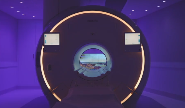
A Pelvis MRI scan is an imaging test used to generate detailed pictures of the pelvic bones and surrounding tissues. These scans are often ordered for patients who are experiencing unexplained hip pain or other issues in the pelvic area. The patient will lie on their back on a platform that moves through the centre of the machine, which uses a strong magnetic field and radio waves to create cross-sectional images of the pelvis. The resulting images can assist doctors in making an accurate diagnosis and determining appropriate treatment options. Pelvic MRI scans are non-invasive and usually completed within 30 minutes to an hour. Patients do not experience any discomfort during the scan, though those with a fear of enclosed spaces may feel anxious in the narrow MRI scanner. However, sedatives can be given to help these patients relax during their scan appointment. This kind of MRI scan differs from other imaging tests, such as X-rays and CT scans, because it does not use ionizing radiation. As a result, there is no risk of exposure to harmful radiation with a Pelvis MRI scan.
Why might you need a pelvis MRI?
If you have pain in your pelvis, your doctor may order a pelvic MRI to help determine the source of the pain. Pelvic MRI can also be used to evaluate the cause of urinary or fecal incontinence or to assess pelvic organ prolapse.
Your doctor may also recommend a pelvic MRI if you have the following:
-
Endometriosis: This is a condition in which the tissue that normally lines the inside of your uterus (the endometrium) grows outside of your uterus.
-
Uterine fibroids: These are non-cancerous growths that develop in or on the wall of the uterus.
-
Ovarian cysts: These are fluid-filled sacs or pockets that can form on or around the ovaries.
-
Pelvic inflammatory disease: This is an infection of the female reproductive organs that can lead to scarring and damage.
-
Ectopic pregnancy: This is a pregnancy that occurs when the fertilized egg implants outside of the uterus, usually in one of the fallopian tubes.
-
Miscarriage: This is the spontaneous pregnancy loss before the 20th week.
Your doctor may also use magnetic resonance imaging MRI for pelvic to guide biopsies or other procedures such as needle aspiration or tissue ablation. A biopsy is a procedure that removes a small tissue sample from the body for testing. Needle aspiration is a procedure that involves extracting fluid or cells from a cyst or mass with a needle. Tissue ablation is a procedure that destroys abnormal tissue with heat, cold, or electrical energy.
How to prepare for your pelvis MRI?
If you have been scheduled for a pelvic MRI, it is important to take steps to prepare for the procedure. Firstly, inform your doctor about any metal implants or objects in your body, as these can interfere with magnetic resonance imaging. Be sure to remove all jewellery and wear comfortable clothing without any metal accents. Additionally, if you wear hearing aids or other devices, ask your doctor if it is safe to leave them in during the scan. Continue eating and engaging in your usual activities leading up to the MRI – no special physical preparation is necessary. Moreover, a creatinine blood test may be performed for some patients to ensure they do not have kidney problems, which can affect the image quality.
What To Expect from an MRI scan of your Pelvis
During the Scan
If your doctor has recommended an MRI scan of your pelvis, here is what you can expect. At the appointment, you will be asked to change into a hospital gown and remove any metal objects (such as jewellery) that could interfere with the scan. The MRI machine is an enclosed space, but the technician will remain in constant communication with you during the procedure. Your referring physician will have already provided information about why the scan is necessary and any specific instructions for preparation. In some cases, a radiographer may use a contrast dye to provide better imaging of various structures within your pelvic area. This contrast material, usually gadolinium, may be administered through an IV line or as a drink beforehand. Don’t be surprised if you feel slightly tingling as the contrast material enters your bloodstream, but this should only last briefly. During the scan itself, you will lie on a sliding table that moves in and out of the MRI machine as it captures images of your pelvic region and any potential abnormalities within its soft tissues.
Moreover, you will hear a loud banging noise coming from the machine – this is perfectly normal and is simply the sound of the magnetic field aligning. You will be given earplugs or headphones to help muffle this noise.
After the Scan
Since the MRI scan is a non-invasive procedure, you can typically resume your usual activities immediately afterwards. But if you receive contrast material, a radiographer will monitor you for any allergic reaction. Gadolinium is the most common type of contrast agent used in MRIs, and while it does have a shallow risk of causing an allergic reaction, your doctor will want to be sure that you don’t have any adverse reaction before releasing you. If everything goes well, you should be able to return home and resume your normal activities without any problem.
What are the benefits of an MRI scan of the pelvis?
There are many benefits of having a magnetic resonance imaging (MRI) scan of the pelvis. These benefits include:
No Exposure to Ionizing Radiation
A pelvis MRI does not use ionizing radiation, unlike other imaging modalities such as X-rays or CT scans. It means there is no risk of exposure to harmful radiation for the patient.
High Contrast Resolution
MRI images have a very high contrast resolution. It means we can clearly visualize different tissues within the pelvis on the scan.
Excellent Soft Tissue Detail
MRI provides excellent detail of soft tissues within the pelvis. It is especially useful for diagnosing problems with the muscles, tendons, and ligaments.
No Sedation Required
Not all the time, a pelvis MRI can be performed without the need for sedation. But if the patient is Claustrophobic, mild sedation may be used to help them relax during the scan.


